
Bone Grafting: Step-by-Step Surgical Breakdown
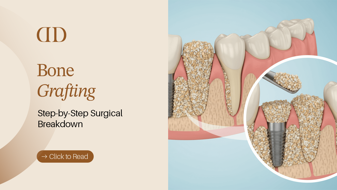

A bone grafting procedure is a minor oral surgery that rebuilds thinned or lost jawbone volume by placing natural or synthetic bone material into areas with bone deficiency. By regenerating solid structural support, bone grafting for dental implants ensures that artificial tooth roots achieve permanent stability and long-term clinical success.
When a tooth is lost or extracted, the surrounding jawbone naturally begins to shrink losing up to 25% of its width within the first year alone. For patients planning a permanent smile reconstruction, finding out you have jawbone loss can feel like an unexpected setback. However, bone regeneration technology in modern dentistry makes it entirely possible to rebuild a firm, healthy foundation for a lasting replacement tooth. At Darya Dental Clinic, located in the vibrant hub of Ataşehir, Istanbul (conveniently close to Ümraniye), our oral surgery team routinely restores severe bone deficiencies, turning complex implant cases into predictable, lifelong success stories.
What Is Bone Grafting?
A dental bone graft is a specialized procedure used in oral and maxillofacial surgery to augment or rebuild jawbone volume that has degraded due to tooth loss, trauma, or gum disease. During the procedure, a specialized graft material is meticulously placed into the deficient site, serving as a biological scaffold. Over a period of several months, your body replaces this matrix with natural, living bone cells through a physiological process called osseointegration and bone regeneration.
A dental bone graft provides a regenerative scaffold that stimulates your body to deposit new, dense jawbone tissue where volume was previously lost. This structural foundation is critical for supporting dental implants and preserving natural facial geometry over a lifetime.
Why Bone Grafting Is Performed
The primary objective of bone grafting in restorative dentistry is to create sufficient jawbone support and structural depth. Dental implants require a specific volume of surrounding bone tissue typically at least 1.5 to 2 millimeters of bone thickness on all sides to withstand the vertical and lateral chewing forces of daily life. When bone volume is insufficient, inserting an implant risks damaging adjacent nerve canals, penetrating sinus cavities, or suffering premature mechanical failure.
Furthermore, jaw bone grafting protects the integrity of your facial aesthetics. Without structural support from internal bone density, the cheeks and lips gradually collapse inward, creating a prematurely aged appearance.
When Bone Grafting Becomes Necessary
Bone grafting for dental implants becomes essential under several specific clinical circumstances:
- Inadequate Alveolar Height or Width: When a 3D Cone Beam Computed Tomography (CBCT dental imaging) scan confirms that the jawbone lacks the height or width required to anchor a standard-sized implant screw.
- Immediate Post-Extraction Protection: Immediately after a tooth extraction, preventing the surrounding socket walls from collapsing.
- Anatomical Proximity: In the upper jaw, where natural sinus pneumatization (expansion of the sinus cavity downward) leaves too little bone height beneath the maxillary sinus floor.
- Severe Periodontal Damage: When advanced gum disease treatment reveals deep osseous defects around remaining teeth.
Common Causes Of Jaw Bone Loss
Understanding why bone loss occurs helps highlight why targeted intervention is so valuable. According to published clinical studies archived in the National Center for Biotechnology Information (NCBI), jawbone degradation is primarily driven by:
- Lack of Functional Stimulation: Natural teeth transfer biting pressure through their roots directly into the alveolar bone. When a tooth is removed, this mechanical stimulation ceases, signalling the body to reabsorb unneeded bone minerals.
- Untreated Periodontal Disease: Bacterial infections in the gums break down soft tissue and erode the underlying hard tissue matrix supporting the teeth.
- Physical Trauma: Facial injuries, jaw fractures, or traumatic impacts during sports can shatter bone structures or sever vascular support, leading to localized necrosis and volume loss.
- Long-Term Removable Denture Wear: Traditional dentures rest on top of the gums without stimulating the underlying bone, gradually accelerating jaw bone loss over years of wear.
Who Needs Bone Grafting Before Dental Implants?
Not every patient receiving an artificial tooth root requires additional bone preparation. However, a significant portion of international patients traveling for a dental bone graft in Istanbul present with subtle or severe bone deficits. Evaluating who needs bone augmentation is the first step toward a predictable, long-lasting treatment outcome.
Premium Dental Care in Turkey
Expert treatments with high-end technology. Get your personalized quote today via WhatsApp.
Bone Loss After Tooth Extraction
Following a routine extraction, the surrounding socket undergoes rapid structural changes. Without intervention, the socket walls naturally resorb. Up to 40% to 60% of the socket height and width can erode within three to six months post-extraction. Patients who delayed getting a replacement tooth right away often require a tooth bone graft to restore the space before an implant can be safely anchored.
Periodontal Disease
Advanced periodontitis is one of the leading causes of missing teeth and structural bone deficiency worldwide. As anaerobic bacteria colonize deep below the gum line, the immune system responds with inflammation that inadvertently breaks down the surrounding alveolar ridge. Patients undergoing periodontal rehabilitation frequently require a periodontal bone graft to rebuild deep structural pockets and stabilize the jaw framework.

Want trusted dental advice in your Google AI answers?
Add Darya Dental Turkey as a Preferred Source on Google to make our dental guides easier to find in your search results and AI Overviews.
Long-Term Missing Teeth
When a gap in the mouth goes unaddressed for years, the localized jawbone progressively thins into a narrow, knife-edge ridge. Attempting to place a standard implant into a thinned ridge without pre-surgical ridge augmentation can result in thread exposure, persistent infection, or implant rejection.
Trauma Or Infection
Severe root canal failures, chronic apical cysts, or direct physical trauma often leave hollowed-out defects in the jawbone. Rebuilding these damaged areas requires structured guided bone regeneration (GBR) techniques to re-establish clean, dense bone before placing long-term restorations such as All-on-4 dental implants or single crowns.
Types Of Dental Bone Grafting Procedures
Depending on the exact anatomical location, defect shape, and severity of bone loss, our oral surgery team at Darya Dental Clinic utilizes distinct, highly targeted surgical methods:
Socket Preservation
Also known as alveolar ridge preservation, this procedure is performed immediately at the time of a tooth extraction. As soon as the natural root is removed, the open socket is cleansed and packed with high-grade bone graft material, then sealed with a protective membrane or sutures. This proactive step prevents the socket walls from collapsing and preserves the natural contour for future implant placement.
Ridge Augmentation
When a missing tooth space has been left open for months or years, the jawbone shrinks in width and height. Ridge augmentation involves making a precise incision along the gumline, lifting the soft tissue, and packing graft material directly onto the narrow bone ridge to restore its original volume and natural width.
Sinus Lift Bone Grafting
In the posterior upper jaw (the molar and premolar regions), the bone is naturally thinner, and the maxillary sinus floor sits just above the tooth roots. If teeth in this area have been missing for an extended period, the sinus cavity expands downward, leaving inadequate bone height for an implant. A sinus lift procedure gently lifts the sinus membrane upward and fills the underlying space with bone material, creating safe depth for implant placement.
Guided Bone Regeneration (GBR)
Guided bone regeneration (GBR) is a advanced technique utilized for localized bone defects around teeth or implants. GBR combines specialized graft material with a biocompatible barrier membrane (resorbable or non-resorbable). This membrane acts as a protective barrier, preventing fast-growing soft tissue cells from invading the surgical site while allowing slower-growing hard tissue bone cells to regenerate undisturbed.
Bone Grafting Procedure Comparison
The table below outlines the primary indications, biological objectives, and average healing windows associated with the main surgical approaches:
| Procedure Type | Best For | Clinical Objective | Average Healing Time |
| Socket Preservation | Immediately post-extraction | Prevents socket collapse & ridge shrinkage | 3 to 4 Months |
| Ridge Augmentation | Restoring thin, narrow jaw ridges | Rebuilds bone width and height | 4 to 6 Months |
| Guided Bone Regeneration (GBR) | Localized defects & missing walls | Shields bone growth using barrier membranes | 4 to 6 Months |
| Sinus Lift | Upper posterior jaw volume deficiency | Elevates sinus membrane to create bone depth | 6 to 9 Months |
Step-by-Step Surgical Breakdown
Understanding the sequential phases of bone grafting for dental implants helps alleviate pre-surgical anxiety and prepares you for a smooth, predictable recovery. At Darya Dental Clinic, every surgical intervention is executed with meticulous biological protocols to maximize graft acceptance and long-term implant stability.
1. Clinical Examination and Digital Imaging
The journey begins with an in-depth clinical evaluation supported by high-resolution CBCT dental imaging (3D Cone Beam Computed Tomography). Unlike traditional 2D X-rays, 3D CBCT scans provide a high-precision volumetric map of your jawbone, detailing structural depth, bone density, and the exact spatial location of adjacent nerves and maxillary sinuses. This diagnostic precision ensures that your personalized treatment planning is mathematically calibrated before any incision is made.
2. Treatment Planning
Using advanced digital smile and bone simulation software, our oral surgery team maps out the precise graft boundaries. During this stage, your surgeon selects the optimal graft material and determines whether a concurrent implant placement is possible or if a dedicated healing window is required.
3. Local Anaesthesia
To guarantee a completely pain-free surgical experience, localized anesthesia is carefully administered around the treatment zone. For international patients experiencing heightened dental anxiety or undergoing extensive multi-site jaw bone grafting, conscious sedation options are available to ensure total physical and emotional comfort throughout the procedure.
4. Preparing the Graft Site
Once complete numbness is confirmed, the oral surgeon makes a gentle, micro-precise incision along the gumline to elevate a small flap of soft tissue. This exposes the underlying alveolar bone defect. The receiving bone bed is thoroughly cleansed of any chronic granulation tissue and lightly conditioned (decorticated) to stimulate micro-bleeding, which releases essential growth factors and blood supply required to nourish the incoming graft.
5. Placement of Bone Graft Material
The selected natural or synthetic bone matrix is carefully packed into the deficient site. The surgeon shapes the material to recreate the natural, ideal contours of a healthy jaw ridge, filling deep structural voids or building out narrow bone walls.
6. Membrane Placement When Needed
To protect the newly placed bone matrix, a specialized biocompatible barrier membrane (often made of resorbable collagen) is positioned over the graft. Used extensively in guided bone regeneration (GBR), this membrane acts as a protective shield, preventing fast-growing gum tissue from invading the space while allowing slower-growing bone cells to regenerate uninterrupted.
7. Suturing and Healing
Finally, the soft tissue flap is gently repositioned over the graft and sealed with fine surgical sutures. These sutures protect the site from oral fluids and mechanical disturbance, initiating the vital biological healing phase where your body begins depositing new, living bone tissue.
Step-by-Step Surgical Workflow Summary
The table below outlines the patient experience, duration, and main clinical objectives across each phase of the bone graft procedure:
| Surgical Stage | Clinical Action | Patient Experience | Primary Objective |
| 1. CBCT Scan & Planning | 3D volumetric mapping | Quick, non-invasive digital scan | Evaluates exact bone height, width, and density |
| 2. Anaesthesia | Targeted local numbing | Minor pinch sensation, followed by full numbness | Ensures absolute pain control during surgery |
| 3. Site Preparation | Gentle gum elevation | Pressure sensation only; zero sharp pain | Exposes bone defect and stimulates vascularization |
| 4. Graft Placement | Packing regenerative material | Complete physical comfort | Establishes the structural matrix for new bone |
| 5. Barrier Membrane | Laying protective collagen shield | Unfelt by patient | Prevents soft tissue invasion into graft site |
| 6. Suturing | Soft tissue closure | Non-invasive final step | Protects surgical site and promotes primary healing |
Bone Graft Materials Used In Modern Dentistry
Modern oral surgery utilizes a diverse range of biologically safe, high-grade materials to rebuild lost jawbone. Selecting the ideal graft material depends on the size of the bone defect, patient preference, and the required speed of bone regeneration.
Autograft
An autograft uses living bone harvested directly from another area of the patient’s own body (typically from the chin, ramus, or posterior jaw).
- Advantages: Considered the gold standard in bone regeneration because it contains living osteoblasts (bone-forming cells) and growth factors, eliminating any risk of immune rejection.
- Considerations: Requires a second surgical donor site, which can slightly increase post-operative discomfort.
Allograft
An allograft consists of human donor bone tissue sourced from accredited, highly regulated bone banks. The tissue undergoes rigorous sterilization and demineralization processes to ensure absolute safety and eliminate cellular antigens.
- Advantages: Eliminates the need for a secondary donor site while providing an excellent natural scaffolding that encourages rapid host bone ingrowth.
- Considerations: Absorbs slightly faster than synthetic alternatives, requiring careful surgical containment.
Xenograft
A xenograft is derived from clean, biologically processed animal sources most commonly bovine (cow) or porcine (pig) origin. The organic cellular components are completely removed through high-temperature processing, leaving behind a pure, biologically compatible mineral matrix.
- Advantages: Resorbs very slowly in the body, making it exceptionally effective at maintaining structural volume over long periods (ideal for sinus lift procedures).
- Considerations: Takes longer to fully replace with host bone compared to autografts.
Synthetic Bone Graft Materials
Also known as alloplasts, synthetic bone substitutes are manufactured in laboratories using biocompatible minerals such as calcium phosphate, hydroxyapatite, or beta-tricalcium phosphate.
- Advantages: Completely synthetic and mineral-based, making it an excellent choice for patients with ethical or religious preferences against human or animal-derived materials. Zero risk of disease transmission.
- Considerations: Serves strictly as a structural scaffold (osteoconductive) without inherent cellular growth factors, making it best suited for minor to moderate socket defects.
Healing After Bone Grafting
The biological success of bone grafting relies heavily on the healing process that takes place beneath the surface over the following weeks and months. Knowing what to expect during recovery ensures a comfortable and predictable healing experience.
Healing Timeline
Bone regeneration occurs in distinct physiological phases:
- First 24 to 48 Hours: Blood clot formation stabilizes the graft material within the site. Mild swelling and soft tissue tenderness peak during this window.
- Days 3 to 7: The initial surgical incision closes as soft tissue cells re-attach. Swelling and minor bruising rapidly subside.
- Weeks 2 to 6: Micro-vascular networks grow into the graft material, bringing oxygen and essential nutrients to support new cell growth.
- Months 3 to 6: Osteoblasts gradually replace the biological scaffolding with dense, living natural jawbone tissue a state of mature osseointegration ready for dental implant placement.
What To Expect During Recovery
Post-operative discomfort following a routine dental bone graft is typically mild and easily managed. Most patients describe feeling slight pressure or dull aching rather than sharp pain. Prescribed anti-inflammatory medications and cold compresses applied to the cheek effectively minimize localized swelling. It is completely normal to notice tiny, sand-like granules escaping from the surgical site during the first few days; this is simply excess graft material shedding naturally from the gum margins.
Foods To Eat
Maintaining optimal nutrition without disturbing the healing gums is essential during the initial recovery phase:
- First 3 Days: Stick strictly to cool, soft foods such as smooth yogurts, lukewarm soups, protein shakes, mashed potatoes, scrambled eggs, and smoothies (eaten with a spoon, never a straw).
- Days 4 to 10: Gradually introduce soft solids like well-cooked pasta, tender fish, steamed vegetables, and soft rice.
- Hydration: Drink plenty of water, but avoid extremely hot liquids (like scalding coffee or tea) for the first 48 hours, as heat can disrupt clot formation.
Activities To Avoid
Protecting the delicate graft site requires temporary lifestyle modifications:
- Avoid Drinking Through a Straw: The suction pressure created in the mouth can dislodge the blood clot and dislocate the graft granules.
- No Smoking or Vaping: Nicotine constricts blood supply to the gums, drastically increasing the risk of graft failure and delayed healing.
- Avoid Strenuous Exercise: Heavy lifting or intense cardiovascular workouts raise blood pressure during the first 3 to 5 days, which can trigger bleeding at the surgical site.
- Do Not Touch the Site: Resist the urge to probe the area with your tongue, fingers, or toothpicks.
Bone Grafting Cost
Navigating the financial investment for a dental bone graft requires understanding that oral reconstruction is never a standard, off-the-shelf product. Because jawbone degradation varies significantly from patient to patient, the bone graft dental cost is always calculated based on the precise anatomical complexity of the defect, the volume of material required, and the specific surgical technique used.
Rather than providing arbitrary fixed numbers that may mislead patients, a transparent clinical practice evaluates the exact scope of treatment using 3D CBCT diagnostic imaging.
Factors Affecting Bone Grafting Cost
Several key biological and surgical variables dictate the overall periodontal bone graft cost and structural preparation fees:
- Type and Source of Graft Material: High-grade xenografts and synthetic matrices carry different processing costs compared to complex autografts, which require a secondary surgical harvesting site.
- Volume of Bone Material Required: Filling a small post-extraction socket (socket preservation) requires significantly less material than rebuilding a multi-tooth, thinned jaw ridge (ridge augmentation).
- Need for Specialized Barrier Membranes: Incorporating resorbable collagen membranes for guided bone regeneration (GBR) adds to the overall surgical material expense.
- Concurrent Surgical Procedures: Combining a bone graft with a sinus lift procedure or complex extractions alters the scope of the surgery.
- Geographic and Operational Economics: Receiving a dental bone graft in Istanbul offers substantial financial savings compared to Western Europe or the US, driven by lower operational overheads and favorable exchange rates without compromising European-standard medical devices or clinical expertise.
Why Every Patient Requires a Personalised Treatment Plan
Two patients seeking an implant for the exact same tooth location may present with completely different underlying bone densities. One may have adequate native bone requiring only minor socket preservation, while another who lost their tooth years prior may require extensive jaw bone grafting and a sinus lift before an implant can be anchored safely.
At Darya Dental Clinic, we believe in absolute financial transparency. Following a remote or in-person 3D CBCT evaluation, our clinical team provides an itemized, all-inclusive treatment plan outlining every phase of your care ensuring zero unexpected fees during your journey.
Why Choose Darya Dental Turkey For Bone Grafting?
When traveling internationally for complex oral surgery, selecting an elite clinic that combines advanced diagnostic technology with deep surgical expertise is paramount. Darya Dental Clinic stands as a premier center for restorative dentistry and maxillofacial rehabilitation in Turkey.
Digital Diagnosis
We eliminate guesswork through state-of-the-art 3D Cone Beam Computed Tomography (CBCT). This technology allows our surgeons to analyze your jawbone density, bone height, and nerve pathways with micrometer precision, ensuring highly predictable outcomes.
Experienced Implant Team
Our team consists of highly trained oral and maxillofacial specialists with extensive experience in handling complex bone deficiencies, failed prior implants, and full-mouth rehabilitations.
European Standard Materials
We exclusively utilize internationally certified, top-tier biocompatible graft materials and barrier membranes imported from leading European and Swiss manufacturers, guaranteeing optimal cellular integration and long-term safety.
Personalised Treatment Planning
Every patient receives a tailored treatment roadmap designed around their specific anatomical needs, medical history, and aesthetic goals balancing clinical efficiency with long-term durability.
Located In Ataşehir, Istanbul
Our modern clinic is situated in Ataşehir, a prestigious, contemporary district on the Asian side of Istanbul. It offers a calm, premium environment for medical recovery, conveniently accessible and close to neighboring districts like Ümraniye.
Dedicated International Patient Coordination
From transparent remote consultations and multi-lingual patient hosts to luxury airport-clinic transfers, we ensure your medical journey to Turkey is comfortable, seamless, and completely stress-free.
Frequently Asked Questions
1. What is bone grafting in dentistry?
A dental bone graft is a specialized surgical procedure that rebuilds lost or thinned jawbone tissue by placing natural or synthetic bone material into deficient areas, creating a stable foundation for dental implants or natural tooth support.
2. Is bone grafting necessary before a dental implant?
Bone grafting is necessary if your jawbone lacks the height, width, or density required to firmly anchor an implant screw. Without adequate bone support, implants risk failure, nerve damage, or sinus penetration.
3. Does bone grafting hurt during or after surgery?
The procedure itself is completely painless as it is performed under advanced local anesthesia or conscious sedation. Post-surgical recovery involves mild swelling and dull aching for 3 to 5 days, which is easily managed with prescribed anti-inflammatory medications.
4. How long does it take to heal after a dental bone graft?
Initial soft tissue healing occurs within 7 to 14 days. Complete bone regeneration and osseointegration where the graft fully matures into solid natural bone typically takes between 3 to 6 months before an implant can be permanently loaded.
5. Can dental implants be placed immediately with a bone graft?
Yes, in cases of mild bone deficiency or immediate socket preservation, an implant screw can often be placed during the exact same surgical appointment as the bone graft. However, severe bone loss requires a separate healing window first.
6. What foods can I eat after bone grafting surgery?
For the first 3 to 5 days, stick strictly to cool, soft foods such as yogurts, lukewarm smooth soups, mashed potatoes, protein shakes, and scrambled eggs. Avoid hot liquids, crunchy, spicy, or seed-filled foods that could irritate the surgical site.
7. What are the risks or main causes of bone graft failure?
While dental bone grafts have a success rate exceeding 95%, potential risks include localized infection, graft rejection, or premature membrane exposure. Smoking, poor oral hygiene, and uncontrolled systemic conditions (like unmanaged diabetes) significantly increase the risk of failure.
8. How much does a dental bone graft cost in Istanbul, Turkey?
The cost depends on the amount of graft material required and the complexity of the procedure (such as concurrent sinus lifts). However, receiving treatment at Darya Dental Clinic in Istanbul generally costs 60% to 70% less than in the UK or US while utilizing identical European-standard materials.
9. Is bone grafting safe for long-term health?
Yes, dental bone grafting is an exceptionally safe, routinely performed oral surgery. The materials used are completely sterile, biocompatible, and engineered to integrate naturally with your body’s cellular framework without causing systemic harm.
10. Can I get a bone graft done immediately after a tooth extraction?
Yes, this procedure is called socket preservation or alveolar ridge preservation. Placing graft material into the open socket immediately after tooth extraction prevents up to 60% of the natural bone collapse that typically occurs during the first year of tooth loss.
Rebuild Your Smile Foundation at Darya Dental Clinic
A lifetime of confident smiling and comfortable chewing begins with a healthy, solid structural foundation. If you have been told you suffer from jawbone loss or need bone preparation before getting dental implants in Turkey, our expert surgical team is here to guide you every step of the way.
At Darya Dental Clinic in Ataşehir, Istanbul (easily accessible from Ümraniye and international transport hubs), we combine digital 3D diagnostics, European-standard biocompatible materials, and compassionate care to make your treatment predictable and stress-free.
Take the first step toward restoring your smile today. Contact our international patient team to upload your dental X-rays or request a free, non-binding digital consultation and personalized treatment plan.
