Routine dental X-rays are a cornerstone of modern dental care, providing critical insights into oral health that cannot be gained through visual examination alone. These imaging tools allow dentists to detect issues early, prevent complications, and tailor treatments to individual needs. While some patients may question the necessity or safety of X-rays, their benefits in maintaining dental health are well-documented. This article explores the importance of routine dental X-rays, their role in diagnosis and treatment, and how they contribute to long-term oral health.
What Are Dental X-Rays?
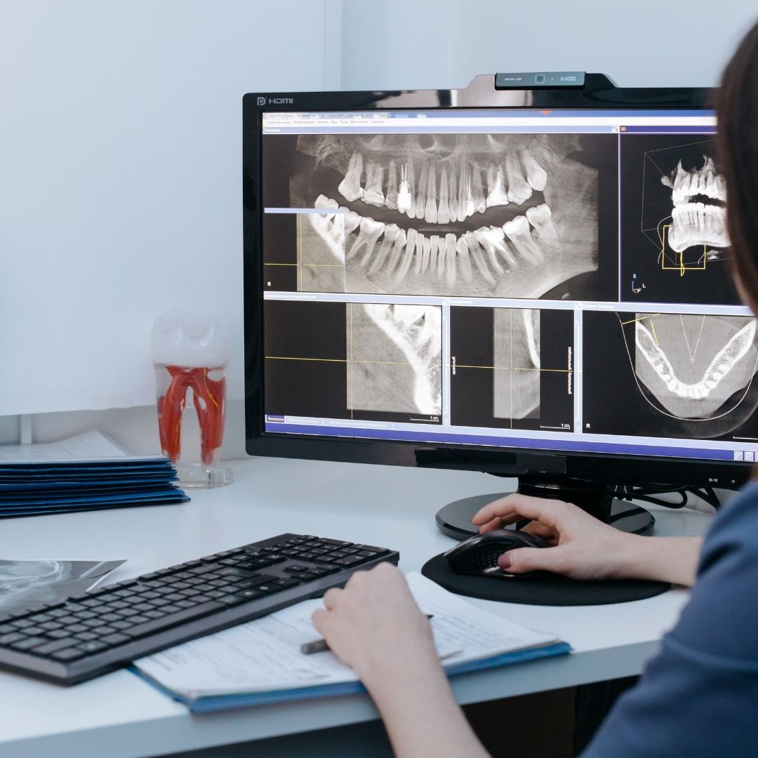
Dental X-rays, also known as radiographs, are images of the teeth, bones, and surrounding soft tissues created using low levels of radiation. They reveal structures invisible to the naked eye, such as tooth roots, bone density, and areas between teeth. Common types of dental X-rays include:
-
Bitewing X-rays: Show the upper and lower back teeth, used to detect cavities between teeth and assess bone levels.
-
Periapical X-rays: Focus on one or two teeth, capturing the entire tooth from crown to root and surrounding bone.
-
Panoramic X-rays: Provide a broad view of the entire mouth, including all teeth, jaws, and sinuses, often used for orthodontics or wisdom teeth evaluation.
-
Cone Beam CT: A 3D imaging technique for complex cases, such as implants or TMJ disorders.
Routine X-rays are typically performed during dental checkups, with frequency depending on age, oral health, and risk factors. The American Dental Association (ADA) recommends X-rays every 1–2 years for adults with good oral health, and more frequently for those with higher risk.
The Role of X-Rays in Dental Care
1. Early Detection of Cavities
Cavities often develop in areas not visible during a visual exam, such as between teeth or beneath fillings. Bitewing X-rays are particularly effective at identifying these hidden caries. Early detection allows for minimally invasive treatments, like small fillings, rather than root canals or extractions. A 2020 study in Caries Research found that X-rays detected 40% more interproximal cavities (between teeth) than clinical exams alone.
2. Identifying Tooth Decay and Damage
X-rays reveal decay that weakens tooth structure, as well as cracks, fractures, or wear not apparent on the surface. For example, periapical X-rays can detect decay extending to the tooth root or abscesses, which may cause pain or infection if untreated. This is critical for saving teeth and preventing systemic complications, as untreated dental infections can spread to the bloodstream.
3. Monitoring Bone Health
The jawbone supports teeth, and bone loss is a hallmark of periodontal (gum) disease. X-rays show bone density and structure, helping dentists diagnose early-stage gum disease or monitor its progression. Panoramic X-rays are especially useful for assessing bone health in older adults or those with a history of gum issues. According to the CDC, 47% of adults over 30 have some form of periodontal disease, making routine X-rays essential for early intervention.
4. Evaluating Tooth Development and Alignment
For children and adolescents, X-rays track tooth development, identifying issues like impacted teeth, crowding, or abnormal eruption. In adults, panoramic X-rays help assess wisdom teeth positioning, which can cause pain or damage if impacted. Orthodontic treatments, such as braces or aligners, often rely on X-rays to plan precise tooth movements.
5. Detecting Abnormalities
X-rays can identify cysts, tumors, or other abnormalities in the jaw or soft tissues. While rare, these conditions can be serious, and early detection through routine X-rays improves outcomes. For example, panoramic X-rays may reveal signs of oral cancer, which affects over 50,000 Americans annually, according to the Oral Cancer Foundation.
6. Guiding Dental Procedures
X-rays are indispensable for planning treatments like implants, root canals, or extractions. Cone Beam CT scans, for instance, provide 3D images to ensure precise implant placement, reducing complications. Similarly, X-rays guide endodontists during root canals by showing the root canal’s shape and depth.
Why Routine X-Rays Are Necessary
Routine X-rays are proactive, catching problems before they cause symptoms. Many dental issues, like early cavities or bone loss, are asymptomatic in their initial stages. By the time pain or swelling occurs, the condition may require invasive or costly treatments. Routine X-rays enable dentists to:
-
Prevent Progression: Addressing issues early prevents minor problems from becoming major ones.
-
Personalize Care: X-rays inform tailored treatment plans based on individual anatomy and risk factors.
-
Save Costs: Early intervention is often less expensive than treating advanced issues like tooth loss or gum surgery.
-
Protect Overall Health: Oral health is linked to systemic conditions, such as heart disease and diabetes. X-rays help maintain oral health, reducing these risks.
For high-risk patients—those with a history of cavities, gum disease, or systemic conditions like diabetes—routine X-rays are even more critical. The ADA notes that diabetic patients are twice as likely to develop periodontal disease, underscoring the need for regular imaging.
Safety of Dental X-Rays
Concerns about radiation exposure often deter patients from agreeing to X-rays. However, modern dental X-rays use minimal radiation, especially with digital technology, which reduces exposure by up to 90% compared to traditional film X-rays. According to the ADA, a full set of dental X-rays exposes patients to about 0.005 millisieverts (mSv) of radiation, equivalent to a few hours of natural background radiation from the environment.
Protective measures further enhance safety:
-
Lead Aprons and Thyroid Collars: These shield the body and sensitive areas from scattered radiation.
-
Digital X-Rays: These require less radiation and produce instant, high-quality images.
-
Targeted Imaging: Dentists use the smallest field of view necessary to minimize exposure.
For context, a single dental X-ray exposes you to less radiation than a cross-country flight (0.01–0.02 mSv). Pregnant patients should inform their dentist, as X-rays are typically deferred unless essential, with extra precautions like double lead shielding.
Who Needs Routine X-Rays?
The frequency and type of X-rays depend on individual factors:
-
Children: Need more frequent X-rays (every 6–12 months) to monitor developing teeth and jaws.
-
Adults with Good Oral Health: May need bitewing X-rays every 1–2 years and panoramic X-rays every 3–5 years.
-
High-Risk Adults: Those with gum disease, frequent cavities, or medical conditions may require X-rays every 6–12 months.
-
New Patients: Often receive a full-mouth series of X-rays to establish a baseline for future comparisons.
Dentists assess risk factors like oral hygiene habits, diet, and medical history to determine X-ray schedules. Patients with braces, implants, or recent dental trauma may need additional imaging.
Addressing Common Concerns
1. Cost
X-rays are often covered by dental insurance as part of preventive care, but out-of-pocket costs can range from $25–$200, depending on the type and number of images. Discuss costs with your dentist upfront to avoid surprises.
2. Overuse
Some patients worry about unnecessary X-rays. Reputable dentists follow ADA guidelines, only recommending X-rays when clinically justified. If concerned, ask your dentist to explain the rationale for each image.
3. Discomfort
X-rays are non-invasive, but biting on sensor holders can be uncomfortable for some. Modern digital sensors are smaller and more comfortable, and dentists can adjust techniques for sensitive patients.
4. Alternatives
While visual exams and intraoral cameras provide some information, they cannot detect subsurface issues like X-rays. No alternative matches their diagnostic precision for hidden problems.
Maximizing the Benefits of X-Rays
To ensure X-rays are effective:
-
Share Your History: Inform your dentist of past X-rays, medical conditions, or pregnancy to tailor imaging.
-
Follow Recommendations: Adhere to the suggested X-ray schedule to catch issues early.
-
Choose a Modern Practice: Opt for offices with digital X-ray technology for lower radiation and better image quality.
-
Maintain Oral Hygiene: Brushing, flossing, and regular cleanings complement X-rays by reducing the likelihood of issues.
The Broader Impact of Routine X-Rays
Routine X-rays contribute to a proactive approach to dental care, aligning with the shift toward preventive medicine. By identifying issues before they escalate, X-rays reduce the need for invasive procedures, lower healthcare costs, and improve quality of life. They also support interdisciplinary care, as dentists share X-ray findings with orthodontists, oral surgeons, or physicians when needed.
For example, X-rays revealing bone loss may prompt a referral to a periodontist, while abnormalities like cysts may require further medical evaluation. This collaborative approach ensures comprehensive care, especially for patients with complex health needs.
Conclusion
Routine dental X-rays are a vital tool in maintaining oral health, offering insights that visual exams cannot provide. From detecting hidden cavities to guiding complex treatments, X-rays enable early intervention, personalized care, and better outcomes. While concerns about radiation or cost are valid, modern technology and professional guidelines ensure safety and necessity. By incorporating routine X-rays into your dental care plan, you invest in a healthier smile and overall well-being, preventing small issues from becoming significant problems.
Frequently Asked Questions (FAQs)
-
How often should I get dental X-rays?
Adults with good oral health typically need X-rays every 1–2 years, while children or high-risk adults may need them every 6–12 months, based on dentist recommendations. -
Are dental X-rays safe?
Yes, modern digital X-rays use minimal radiation, equivalent to everyday environmental exposure. Lead aprons and targeted imaging further enhance safety. -
Can I refuse dental X-rays?
You can refuse X-rays, but this may limit your dentist’s ability to detect hidden issues, potentially leading to untreated problems. -
Why can’t a visual exam replace X-rays?
Visual exams cannot detect subsurface issues like cavities between teeth, root decay, or bone loss, which X-rays reveal clearly. -
Are dental X-rays covered by insurance?
Most dental insurance plans cover routine X-rays as part of preventive care, but coverage varies. Check with your provider for details.